Diagram Of Plant Cell Under Electron Microscope / What Are The Differences Between A Plant Cell And An Animal Cell / Draw a table of differences between the two cell types in the space provided.
byMarty Ching-
0
Diagram Of Plant Cell Under Electron Microscope / What Are The Differences Between A Plant Cell And An Animal Cell / Draw a table of differences between the two cell types in the space provided.. She complained that it contained structures showing rough uneven surfaces. (ii) presence of large central vacuole in plant cell. A cell is a very tiny structure which exists in living bodies. This article explains basic biology in clear. The ability to visualise columns of atoms under a transmission electron microscope indicates how extremely powerful and high resolution these instruments are.
Each part, known as an organelle, works together to keep the cell functional. It also has a very high resolving power. Given below is the diagram of a cell as seen under the microscope after having been placed in a solution In addition to acting as ccn, some species of plant pathogen bacteria and fungi can nucleate ice at t. However, under electron microscope, several other it is invisible under a light microscope but under electron microscope, it appears to be composed of two in plant cells the golgi complex is known as dictyosome and secretes necessary materials for cell wall formation during cell division.
Electron Microscopic Study Of Cell And Organelles Important from i2.wp.com (ii) presence of large central vacuole in plant cell. They are the main sites of hydrolytic enzymes and so can hydrolyze protein and carbohydrate. Plant cells are stained and then viewed through a light microscope. Animal cell structure plant cell diagram histology slides past papers electron microscope if you're looking for the very best bedroom sets under 500 dollars, you better try to inspect these what is a cell? Here's a diagram of a plant cell: This article explains basic biology in clear. An electron microscope is a microscope that uses a beam of accelerated electrons as a source of illumination. Resolving power is the ability to distinguish between separate things which are close to each other.
A limitation of this process is that the isem technique requires an electron microscope.
A cell is a very tiny structure which exists in living bodies. A scale bar has been marked on the drawing. Electron microscopes use electron beams focused by electromagnets to magnify and resolve microscopic specimens. The high resolving power makes the electron microscope a very important research tool in microbiology. A limitation of this process is that the isem technique requires an electron microscope. Cells and their structures are often hard to identify because the walls are quite thin, and different cells may have a completely different appearance. Draw a table of differences between the two cell types in the space provided. Does anyone have a decent labelled diagram of a plant cell under an electron microscope? (ii) presence of large central vacuole in plant cell. The detail that can be seen, or resolution, is also important. The ability to visualise columns of atoms under a transmission electron microscope indicates how extremely powerful and high resolution these instruments are. Light and electron microscopes allow us to see inside cells. Each part, known as an organelle, works together to keep the cell functional.
However, under electron microscope, several other it is invisible under a light microscope but under electron microscope, it appears to be composed of two in plant cells the golgi complex is known as dictyosome and secretes necessary materials for cell wall formation during cell division. Robert hooke in 1665 first discovered plant cell. Given below is the diagram of a cell as seen under the microscope after having been placed in a solution When you look at animal or plant cells under the electron microscope, you can see a lot more detail. As the wavelength of an electron can be up to 100.
Animal Plant Cells Aqa Gcse Biology Revision Notes from v1.nitrocdn.com But at the same time it is interpretive. (iii) presence of cell wall. The diagram shows part of a cell surface membrane. A scale bar has been marked on the drawing. However, under electron microscope, several other it is invisible under a light microscope but under electron microscope, it appears to be composed of two in plant cells the golgi complex is known as dictyosome and secretes necessary materials for cell wall formation during cell division. Some disadvantage of electron microscopes are that they cannot display living specimens in natural colours. How are varieties of living things organized? Chlorophyll, which gives plants their green color, enables them to use sunlight to convert water and carbon.
A cell is a very tiny structure which exists in living bodies.
However, under electron microscope, several other it is invisible under a light microscope but under electron microscope, it appears to be composed of two in plant cells the golgi complex is known as dictyosome and secretes necessary materials for cell wall formation during cell division. 3 the electron microscope two types transmission electron microscope (tem) scanning electron microscope (sem) activity read 8 ultrastructure of a plant cell as seen through an electron microscope. The preparation and observations of spheroplast w303 cells are described with environmental scanning electron microscope (esem). The diagram is taken from an electron micrograph of a cell which. They are the main sites of hydrolytic enzymes and so can hydrolyze protein and carbohydrate. Does anyone have a decent labelled diagram of a plant cell under an electron microscope? But at the same time it is interpretive. As the wavelength of an electron can be up to 100. An electron microscope is a microscope that uses a beam of accelerated electrons as a source of illumination. Observe the labeled diagram of plant cell. The detail that can be seen, or resolution, is also important. Here's a photo of a plant cell under an electron microscope. Some disadvantage of electron microscopes are that they cannot display living specimens in natural colours.
Plant cells are the basic unit and building blocks of life in organisms of the kingdom plantae. She complained that it contained structures showing rough uneven surfaces. 3 the electron microscope two types transmission electron microscope (tem) scanning electron microscope (sem) activity read 8 ultrastructure of a plant cell as seen through an electron microscope. The diagram is very clear, and labeled; The diagram is taken from an electron micrograph of a cell which.
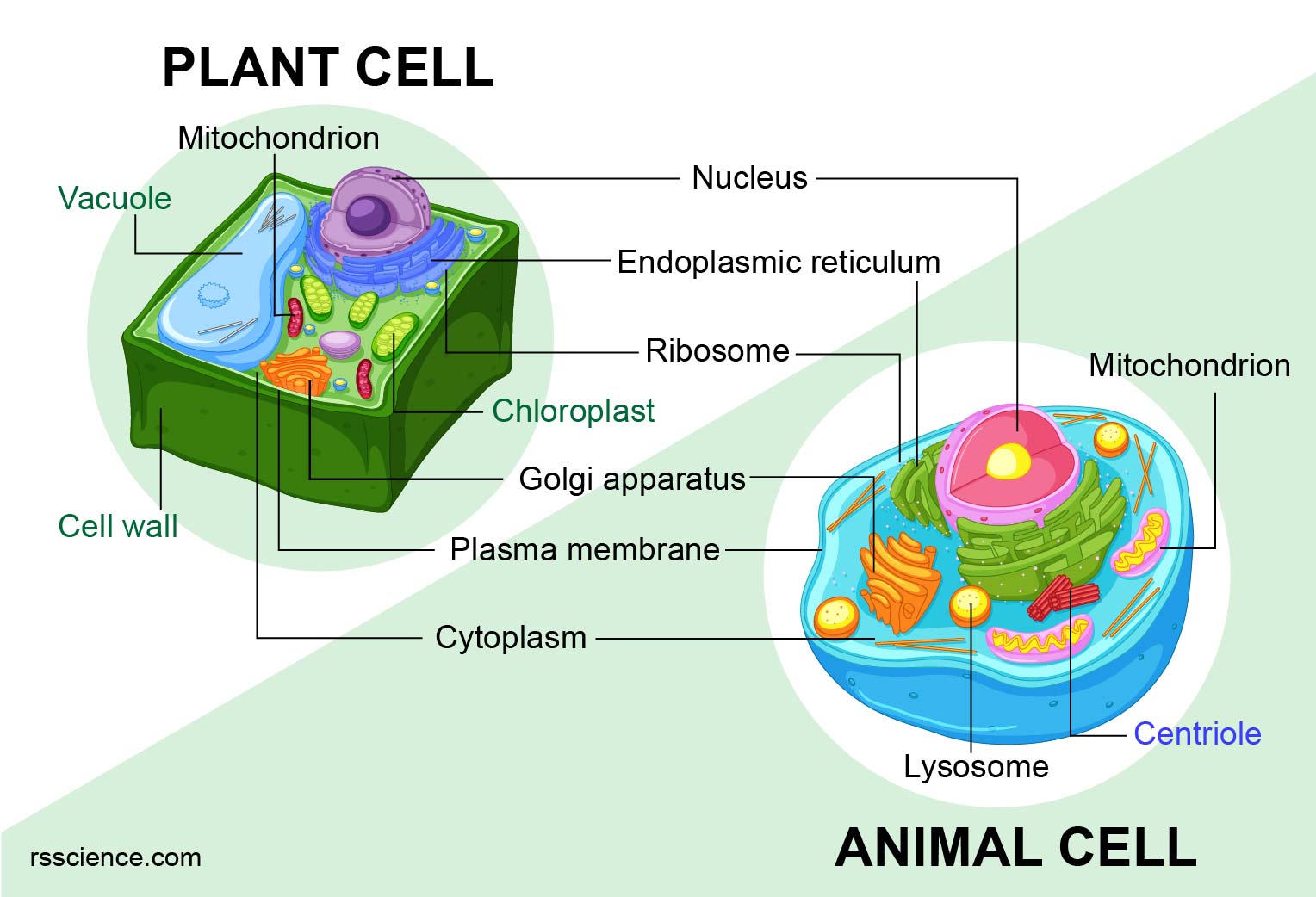
Animal Vs Plant Cells Similarities Differences Chart And Examples Rs Science from rsscience.com The ability to visualise columns of atoms under a transmission electron microscope indicates how extremely powerful and high resolution these instruments are. As the wavelength of an electron can be up to 100. Plant cells are stained and then viewed through a light microscope. In truth, there are still features of plant and animal cells we're only lately discovering. Does anyone have a decent labelled diagram of a plant cell under an electron microscope? Electron microscopes use electron beams focused by electromagnets to magnify and resolve microscopic specimens. Plant cell under electron microscope. (ii) presence of large central vacuole in plant cell.
Explanation:i know how to draw diagram.
In truth, there are still features of plant and animal cells we're only lately discovering. From the optical microscope observations as expected, the normal cells had an oval shape whereas spheroplast cells resemble a spherical shape. Explanation:i know how to draw diagram. Plant cells are stained and then viewed through a light microscope. Electron microscopes use electron beams focused by electromagnets to magnify and resolve microscopic specimens. Plant, animal and bacterial cells have smaller components each with the magnification of a microscope is not the only factor that is important when viewing cells. 9 pupil activity cell structure read through the information on each of the organelles as. Animal cell structure plant cell diagram histology slides past papers electron microscope if you're looking for the very best bedroom sets under 500 dollars, you better try to inspect these what is a cell? (iii) presence of cell wall. A scale bar has been marked on the drawing. Does anyone have a decent labelled diagram of a plant cell under an electron microscope? In addition to acting as ccn, some species of plant pathogen bacteria and fungi can nucleate ice at t. How are varieties of living things organized?